
What is a breast ultrasound scan?
Breast ultrasound is a non-invasive imaging test that uses sound waves to produce detailed images of the internal structure of the breast. This imaging test can determine whether a lump found in the breast during a mammogram (breast X-ray) or physical examination is cancerous or not.
In Singapore, breast ultrasound is widely available and can be done at private clinics, polyclinics, or hospitals. It can be done alongside a mammogram as part of breast cancer screening or if your healthcare provider wants to take a closer look at areas that show abnormalities.
Check out our site to learn more about our health screening services and how they can benefit you.
How does breast ultrasound works
A breast ultrasound uses an ultrasound probe (transducer) that emits high-frequency sound waves to produce detailed images of the breast tissue. During the procedure, an ultrasound technician (sonographer) will apply a clear gel to your breast to help the transducer glide smoothly over the skin.

As the sonographer gently moves the probe over your breast, sound waves bounce off different structures in your breast and return to the probe at different speeds. These reflected sound waves are then converted by a computer into real-time images (sonograms) that can be displayed on a screen.
These real-time images allow the sonographer to thoroughly examine your entire breast and look for abnormalities, such as:
Cysts, which are fluid-filled sacs that aren't cancerous.
Solid masses such as fibroadenomas (non-cancerous lumps) or breast cancer.
Why might I need a breast ultrasound?
Breast ultrasound scans are usually recommended by your healthcare provider after mammography, especially if there are areas on the mammogram results that require further examination.
Here are some conditions where your healthcare provider may recommend a breast ultrasound, such as:
Evaluating a breast lump
This imaging test helps your healthcare provider to determine whether a breast lump is a cyst or a solid mass.
Further examination after a mammogram
If your mammogram reveals an unclear or suspicious area, an ultrasound test can be performed to provide additional detail for more accurate evaluation.
This imaging test is helpful when checking for palpable lumps or changes that are physically felt but not clearly visible on a mammogram.
Women that have dense breast tissue
If you have dense breast tissue, mammograms may not be as effective in detecting lumps. In this case, an ultrasound scan allows a better examination of deeper breast tissues.
Guiding a breast biopsy
As breast ultrasound provides real-time images of the inside of the breast, it can be used to guide healthcare providers to ensure that they take tissue samples from the right place during breast biopsy procedures.
Monitor breast changes
If you have a non-cancerous condition, such as fibroadenoma or a previous breast abnormality, an ultrasound examination can help track changes in size, shape, or appearance over time.
This continuous monitoring helps to ensure that benign conditions remain stable.
Examining other breast abnormalities
Ultrasound is useful for identifying possible infections, abscesses, or blockages in milk ducts that could cause breast pain or abnormal nipple discharge and pinpointing their location.
Alternative breast imaging for pregnant or younger women
While breast ultrasound is usually done after a mammogram, there are some exceptions. An ultrasound can be done before a mammogram for patients who are under 40 years of age, pregnant women, or nursing mothers.
This advantage is because this imaging test is safer, as it uses sound waves instead of ionising radiation, like mammograms do.
Unlike mammography, a breast ultrasound is not usually used for routine breast cancer screening on its own. This disadvantage is due to its potential to miss early signs of certain cancers, such as microcalcifications, which are small white spots or flecks that are only visible on mammograms.
However, breast ultrasound is an important diagnostic tool that can complement other imaging tests such as mammography or MRI in diagnosing and monitoring various breast conditions.
If you have concerns regarding breast lumps or other breast-related conditions, request an appointment with Thomson Medical. Our specialists can assist with further diagnoses, including a breast ultrasound scan to determine the underlying causes and provide personalised care.
What to prepare before a breast ultrasound
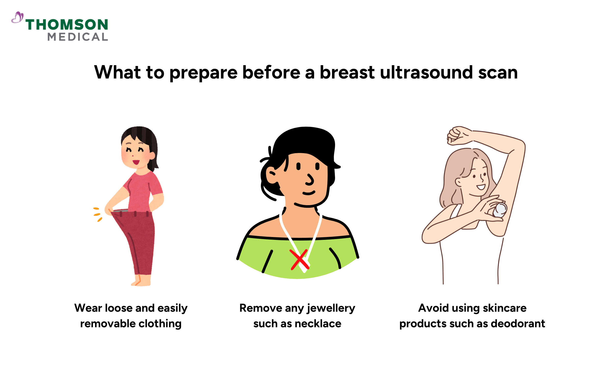
A breast ultrasound scan is a straightforward procedure that only requires minimal preparation prior to the scan, such as:
Wear loose and easily removable clothing, such as a blouse with a skirt or pants, as you will need to undress from the waist up for the procedure.
Avoid using skincare products, such as deodorant, lotions, or powder, on or around your breasts on the day of the scan, as these products can make it difficult to get clear images.
Remove any jewellery from your chest that might interfere with the scan, such as necklaces or body piercings.
If you usually experience breast tenderness during a menstrual cycle, schedule the ultrasound for a time after your menstrual period when your breasts are less sensitive.
What to expect during the scan
Once you’ve completed the preparation steps, the procedure will begin. Here’s what you can expect during the breast ultrasound scan:
You'll be asked to undress from the waist up and remove any jewellery before being given a gown to wear.
Afterwards, you will lie on your back on the examination table and raise your arm above your head.
The sonographer then applies a clear gel on your breast to help the transducer glide smoothly over the skin and make sound waves travel better.
The transducer is then gently pressed against your skin and moved to the area of interest.
The device will then transmit detailed images of the inner breast in real-time so you and your healthcare provider can observe them on-screen during the scan.
After the ultrasound is completed, the sonographer will wipe away any remaining gel from your skin.
The entire procedure typically lasts between 15 and 30 minutes and is painless, allowing you to immediately resume normal activities afterward.
What do the results mean?
After the scan is finished, the sonographer will send the results to a radiologist, a specialist in medical imaging, for analysis. Once the radiologist has reviewed the images, they will share their findings with your healthcare provider.
Your healthcare provider will then contact you to discuss any abnormal findings, ensuring you fully understand your scan result and guide you through any necessary follow-up procedures, such as:
A mammogram for additional detailed imaging.
A biopsy to determine if cancer cells are present.
Regular follow-up ultrasounds to monitor any changes in the breast lump.
Always follow preparation guidelines and discuss any concerns with your doctor to ensure the most accurate results. Request an appointment with us to find out if a breast ultrasound is the right imaging test for you.
Breast ultrasound costs in Singapore
In Singapore, a breast ultrasound can be performed at a hospital, clinic, or diagnostic centre. The cost of this imaging test depends on whether it's performed as a standalone test after a mammogram or as part of a health screening package, as well as the choice of medical facility.
To help cover the cost, you can use up to SGD 300 annually from your MediSave account at select clinics. If you have private insurance, you may need to check with your individual provider to see if a breast ultrasound is covered by your policy.
For detailed fee information and payment options, please consult your healthcare provider directly. Request an appointment with our specialists at Thomson Medical today for a detailed price breakdown and a personalised care plan.
What are the risks of a breast ultrasound?
Breast ultrasound is a safe, non-invasive imaging procedure that uses sound waves instead of ionising radiation. There are no known risks associated with this procedure, although you may experience mild discomfort due to the transducer being pressed against your skin by the sonographer during the procedure.
FAQ
What will an ultrasound of the breast show?
A breast ultrasound can produce detailed images of the internal structure of the breast tissue. Healthcare providers use it to check for lumps in the breast, which can be fluid-filled cysts (which are usually not cancerous) or solid masses such as fibroadenomas (non-cancerous tumours) or potentially cancerous cells.
This imaging test can also be used to detect possible infections, abscesses, or blockages in the milk ducts that could cause breast pain or abnormal nipple discharge.
When is the best time to do a breast ultrasound?
If you experience breast tenderness related to your menstrual cycle, it's advisable to schedule your breast ultrasound shortly after your period ends, when your breasts are less sensitive. This timing ensures a more comfortable procedure and clearer images, as breast tissue tends to be less dense and tender during this time.
Recommended timing for women with menstrual cycles:
Ideal timing is between days 5 and 10 of your cycle (approximately one week after your period begins).
Avoid scheduling just before or during your period, as hormonal fluctuations can cause breast swelling or lumpiness that may affect the clarity of ultrasound images.
For pregnant, breastfeeding, or postmenopausal women:
Ultrasound can be scheduled at any time, as hormonal stability means breast tissue remains consistent, ensuring clear and accurate results.
Can you see cancer on a breast ultrasound?
Yes, a breast ultrasound can identify solid masses within the breast that may indicate cancer. However, this imaging test alone cannot definitively diagnose cancer. If a suspicious mass is detected, additional evaluations such as a biopsy, mammography, or MRI are necessary for further diagnosis.
Which is better, a mammogram or a breast ultrasound?
Neither is universally "better", as they serve different purposes and complement each other. Mammograms are the standard for routine breast cancer screening, as they can detect microcalcifications and other early signs of cancer that ultrasounds might miss.
However, breast ultrasounds can be used if a patient has dense breast tissue and provides additional detail about abnormalities found during mammograms. Ultrasounds are also safer for pregnant women and younger patients, as they use sound waves instead of radiation.
For most patients, the best approach is often a combination of both tests as recommended by your healthcare provider.
Do you get results immediately after a breast ultrasound?
The timing for receiving your ultrasound results varies based on the facility and your doctor's procedures. Results can sometimes be available immediately, especially if there are no identified concerns. Your healthcare provider might discuss the findings right away or shortly after your exam to provide preliminary feedback.
However, in other instances, the results may take longer. Typically, a radiologist reviews the ultrasound images and prepares a detailed report for your doctor, which usually takes between one to three days but occasionally up to a week.
The information provided is intended for general guidance only and should not be considered medical advice. For personalised recommendations based on your medical conditions, request an appointment with Thomson Medical.
For more information, contact us:
Thomson Specialists Paragon (Health Screening)
- Mon - Fri: 8.30am - 5.30pm
- Sat: 8.30am - 12.30pm
Call: 6735 0300
Request a Health Screening