
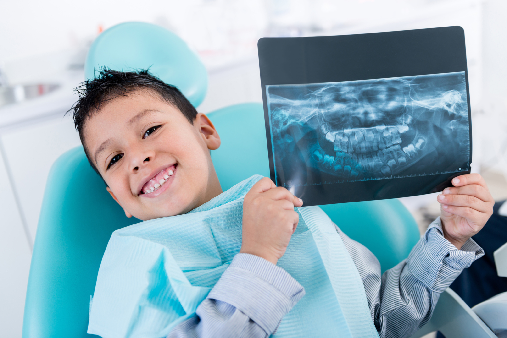
What are dental x-rays?
Dental X-rays, also known as dental radiographs, are images of the teeth and surrounding structures that are captured through the use of X-ray technology. These images are an essential tool in dentistry as they provide valuable information that is not visible during a regular dental examination. Dental X-rays can help dentists diagnose and treat various oral health issues.
What types of dental x-rays are there?
Dental X-rays can be broadly classified into two categories.
Intraoral X-rays are where the X-ray films or receptors are placed inside the mouth. Examples: Bitewing (interproximal) X-rays, periapical X-rays, and occlusal X-rays.
Extraoral X-rays are where the film or receptor is outside the mouth. Examples: Panoramic X-rays, lateral cephalometric X-rays (orthodontic X-rays), and Cone Beam Computed Tomography (CBCT).
They both serve different purposes and are obtained with different techniques.
Intraoral x-rays:
Bitewing (interproximal) X-rays
Purpose: Detect decay between the teeth (interproximal decay) and check for changes in bone density caused by gum disease.
How it is taken: The patient bites down on a special X-ray film holder, and the X-ray machine is aligned to capture images of the upper and lower teeth in a specific area of the mouth.
Periapical X-rays
Purpose: Provide a detailed view of the entire tooth, from the crown to the root, and is used to detect abnormalities in the tooth's root structure and surrounding bone.
How it taken: The X-ray film is placed inside the mouth near the targeted tooth, capturing its entire length as well as the surrounding structures.
Occlusal X-rays
Purpose: Reveal the entire arch of teeth in either the upper or lower jaw. They detect issues such as extra teeth, missing teeth, or abnormalities in tooth development. These X-rays are also useful in assessing the presence and position of unerupted teeth.
How it is taken: The patient bites down on a piece of X-ray film or a digital sensor while the X-ray machine is positioned above or below the mouth. The X-ray machine is adjusted to capture an image of the upper or lower teeth, depending on the area of interest.
Extraoral x-rays:
Panoramic X-rays
Purpose: Offer a broad view of the entire mouth, including the jaw, teeth, sinuses, and nasal area. They are useful for assessing impacted teeth, jaw disorders, and overall oral health.
How it is taken: The patient stands or sits in front of the X-ray machine, and a rotating arm captures a panoramic image as it moves around the head.
Lateral Cephalometric X-rays (Orthodontic X-rays)
Purpose: Help plan orthodontic treatment by providing a side view of the head and jaw, assessing the position and alignment of the teeth.
How it is taken: The patient's head is positioned against a cephalostat, and the X-ray machine captures an image of the entire head.
Cone Beam Computed Tomography (CBCT)
Purpose: Provide a 3D view of the teeth and surrounding structures, offering detailed information for complex dental procedures, such as dental implant placement and oral surgery.
How it is taken: The patient stands or sits in front of the X-ray machine. A rotating arm captures hundreds of images as it moves around the head using a cone-shaped X-ray beam focused on the area of interest.
Are dental X-rays really necessary and why?
Dental X-rays, also known as radiographs, are valuable diagnostic tools that provide information not visible during a regular dental exam. Here are several reasons why dental X-rays are necessary:
Detecting tooth decay:
X-rays can reveal areas of decay between teeth or beneath existing restorations (such as fillings). They help dentists identify cavities in their early stages when they are smaller and easier to treat.
Examining tooth roots:
X-rays allow dentists to evaluate the roots of teeth and surrounding bone structure. This is crucial for diagnosing issues such as infections, abscesses, and abnormalities in the root canal.
Monitoring teeth development:
X-rays are commonly used to track the development of teeth in children and adolescents. They help dentists ensure that teeth are growing properly and identify potential issues early on.
Assessing gum health:
X-rays can reveal the level of bone around your teeth, helping dentists diagnose conditions like periodontal disease. This information is vital for developing an effective treatment plan.
Planning for orthodontic treatment:
X-rays are used in orthodontics to assess the alignment of teeth and jaw structure. This information helps orthodontists plan and execute orthodontic treatments such as braces or aligners.
Detecting impacted teeth:
X-rays are instrumental in identifying impacted teeth, particularly wisdom teeth. Impacted teeth can cause pain, swelling, and damage to adjacent teeth, and X-rays help dentists determine the appropriate course of action.
Evaluating Temporomandibular Joint (TMJ) conditions:
X-rays can assist in diagnosing issues with the jaw joint, such as arthritis or dislocation, which may contribute to pain and dysfunction.
Screening for oral cancer:
X-rays may be used as part of a comprehensive examination to detect abnormalities, tumors, or cysts in the oral cavity.
It is important to note that the frequency of dental X-rays depends on your individual health, age, and risk factors. Dentists follow guidelines to minimise radiation exposure and recommend X-rays when they are necessary. If you have concerns about dental X-rays, discuss them with your dentist, who can provide information about the benefits and risks based on your specific situation.
How much radiation is there in a dental X-ray?
Dental X-rays expose patients to very low levels of radiation. The amount of radiation received during a dental X-ray is measured in millirems (mrem). Here are approximate radiation doses for common dental X-rays:
Bitewing (4 films): Approximately 0.5 mrem
Periapical (2 to 4 films): Approximately 0.3 mrem
Panoramic: Approximately 1 to 3 mrem
Cone Beam CT (CBCT): Approximately 10-20mrem
To put these values into perspective, the average person in the United States is exposed to about 620 mrem of radiation annually from various sources, including natural background radiation, medical procedures, and other environmental factors.
Dental X-rays are designed to minimise radiation exposure. Modern X-ray equipment and techniques, as well as the use of protective aprons and collars, further reduce the radiation dose. Additionally, digital radiography has become common in dental practices, offering advantages such as lower radiation doses compared to traditional film-based X-rays.
It is important to note that the benefits of diagnostic X-rays in dentistry, such as early detection of dental issues and prevention of extensive problems, generally outweigh the minimal risks associated with the low levels of radiation exposure. Dentists follow guidelines established by regulators to ensure that X-rays are used judiciously and when necessary for diagnostic purposes.
If you have concerns about radiation exposure, discuss them with your dentist, who can provide information specific to your situation and address any questions you may have.
Are there situations I should not have a dental X-ray?
While dental X-rays are generally safe, there are situations where it might be advisable to postpone or avoid them. Your dentist will consider various factors when deciding whether to proceed with X-rays, and you should communicate openly about your health and any concerns you may have. Here are some situations where caution or avoidance of dental X-rays may be recommended:
Pregnancy, especially during first trimester:
If you are pregnant, especially during the first trimester, your dentist may avoid routine X-rays unless they are necessary for diagnosis or treatment. In such cases, they will take precautions to minimise radiation exposure to the fetus, such as using lead aprons and thyroid collars.
Recent dental X-rays:
If you have had recent dental X-rays, your dentist may consider the cumulative radiation exposure and decide whether additional X-rays are necessary.
Paediatric patients:
Dentists are particularly cautious with radiation exposure in children. They may use alternative diagnostic methods or limit the number of X-rays, especially for routine check-ups.
Low risk of dental issues:
If you have a low risk of dental problems and no symptoms or issues that warrant X-rays, your dentist may opt to postpone them.
Alternative diagnostic methods:
In some cases, alternative diagnostic methods, such as visual examination, may be sufficient, especially if the suspected issue is localised and does not require detailed imaging.
It is crucial to communicate openly with your dentist about your health, concerns, and any history of radiation exposure. Your dentist will consider your individual circumstances and follow established guidelines to ensure that X-rays are used judiciously and when necessary for diagnostic or treatment purposes.
How much is a dental X-ray in Singapore?
The cost can range from SGD 20 to SGD 300, depending on various factors. Public institutions like polyclinics may charge less, but private clinics often have shorter waiting times.
Can I use CHAS or MediSave for dental X-rays?
For dental X-rays, CHAS subsidies are available, limited to 6 X-rays per calendar year. For CHAS Orange card, there are no subsidies. For CHAS Blue card, you can claim $11 per x-ray. Additional details on CHAS subsidies can be found here.
MediSave typically does not cover general dental treatments, such as dental X-rays, unless they involve surgical procedures deemed medically necessary. Non-surgical dental treatments, including routine check-ups, are not eligible for claims under the MediSave scheme.
FAQ
How often should I get dental X-rays?
The frequency of dental X-rays depends on factors such as your age, oral health, and risk for dental issues. For adults, bitewing X-rays are typically taken every 1-2 years, while full-mouth X-rays (panoramic) may be taken every 3-5 years.
Are dental X-rays safe?
Yes, dental X-rays are generally considered safe. The amount of radiation exposure is minimal, and dentists take precautions such as using lead aprons and collars to protect patients from unnecessary radiation.
Can I have dental X-rays during pregnancy?
While routine X-rays are generally avoided during pregnancy, if there is a pressing dental issue that requires diagnostic imaging, your dentist can take precautions to minimise radiation exposure to the fetus, such as using lead aprons.
Are digital X-rays safer than film?
Yes, digital X-rays typically produce less radiation than traditional film X-rays. Digital technology allows for quality images with lower radiation doses.
Do dental X-rays cause cancer?
The risk of developing cancer from dental X-rays is low. The benefits of early diagnosis and treatment of dental issues generally outweigh the minimal risks associated with the low-dose radiation exposure.
How do I prepare for dental X-rays?
Typically, there is no special preparation. You may be asked to remove jewelry or accessories from your head and neck area, and your dentist will provide protective gear such as lead aprons.
Can I refuse dental X-rays?
You have the right to make informed decisions about your healthcare. If you have concerns about dental X-rays, discuss them with your dentist. They can explain the necessity based on your oral health needs and address any specific concerns you may have.
For more information, contact us:
Thomson Dental Centre
Call: 6255 0770
WhatsApp: 8716 9594
Book an Appointment