
What is an internal auditory meatus MRI scan?
The Internal Auditory Meatus (IAM) is a small bony canal in the skull (temporal bone). It carries nerves and blood vessels from the base of your skull (posterior cranial fossa) to the inner ear, which controls hearing and balance.
An MRI of your IAM is a detailed imaging test that examines these tiny passageways and the surrounding structures. It helps doctors get a clear view of:
The bony passages within your temporal bone
The nerves that carry hearing and balance signals from your inner ear to your brain
Blood vessels that supply the structures within your inner ear
Surrounding structures involved in your hearing and balance, such as the cochlea and vestibular labyrinth
The scan can also detect abnormalities in these areas, such as narrowing, malformations, or tumours.
How is an IAM MRI scan different from a brain MRI?
When you have a regular brain MRI, your doctor gets images of your overall brain structures to look for a wide range of neurological conditions. The main difference between this type of imaging and an IAM MRI lies in the specific area of focus and level of detail.
Here's how an IAM MRI differs from a regular brain MRI:
Focus area
While a regular brain MRI examines your entire brain, an IAM MRI specifically targets the internal auditory canals — the small bony passages connecting your inner ear to your brain.
Image details
While a regular brain MRI might include some visualisation of your inner ear, the IAM MRI provides much more detailed images of this specific area.
This advantage makes it the preferred choice when assessing symptoms like hearing loss, tinnitus, vertigo, or balance issues.
Specialised sequences
An IAM MRI uses special imaging techniques and sequences specifically designed to highlight the delicate structures within the internal auditory canal, such as the hearing, balance, and facial nerves.
Purpose
Regular brain MRIs are used to diagnose a wide range of neurological conditions (like stroke, tumours, or multiple sclerosis), while IAM MRIs focus specifically on conditions related to hearing, balance, and inner ear function, such as:
Vestibular schwannomas (acoustic neuromas)
Causes of unexplained hearing loss
Facial nerve disorders
Inner ear abnormalities
Your doctor will recommend the appropriate type of MRI scan based on your specific symptoms and the conditions they suspect might be causing them.
When is MRI IAM used?
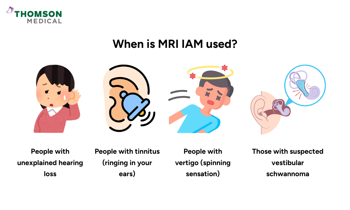
Your doctor might recommend an MRI of your Internal Auditory Meatus (IAM) if they need to examine the structures in your inner ear canal more closely. This specialised test is particularly useful in several situations:
To detect specific conditions
Vestibular schwannomas (small tumours on your hearing and balance nerves)
Meningiomas (tumours that develop from the membranes covering your brain)
Vascular lesions (abnormalities in the blood vessels inside the IAM)
Cystic lesions (fluid-filled sacs that can develop in or near the inner ear and the middle ear)
To evaluate common symptoms
Unexplained hearing loss, especially if it affects only one ear
Tinnitus (ringing in your ears)
Vertigo (spinning sensation) or balance issue
For surgical planning and follow-up
Before surgery to provide detailed images for surgical planning
After surgery, check the results, particularly the following:
Removal of vestibular schwannomas
Cochlear implantation procedures
If you're experiencing ongoing hearing loss, tinnitus, dizziness, or balance issues that aren’t improving or have no clear cause, it’s important to consult a healthcare provider. Request an appointment with Thomson Medical for a comprehensive evaluation.
Our specialists will assess your symptoms and may recommend an IAM MRI to accurately diagnose your condition and develop a personalised treatment plan tailored to your needs.
How to prepare for the test?
Before your IAM MRI, it is important to discuss preparation details with your healthcare provider, as requirements may vary between different testing facilities.
Before your appointment
Your healthcare provider will perform a complete physical examination prior to the MRI test.
Be prepared to discuss your full medical history.
Important information to share with your doctor
It is important for you to share the following information with your doctor:
All medications and supplements you're currently taking
Any medication allergies you have
Any medical implants in your body (such as pacemakers, cochlear implants, or metal plates)
Whether you're pregnant or breastfeeding, as MRI scans with contrast dyes might not be suitable
If you experience claustrophobia or anxiety in confined spaces
Addressing concerns
If you're uncomfortable in small spaces, don't hesitate to mention the issue to your doctor. They can often provide options to help make you more comfortable, such as:
Prescribing a sedative to help you relax during the procedure
Recommending an open MRI if available and appropriate for your needs
Providing other accommodations to ensure your comfort
Every imaging centre may have slightly different guidelines, so follow the specific instructions from your healthcare team.
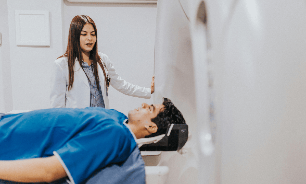
What to expect during the test?
Before your scan begins, you'll be asked to change into a hospital gown. Your MRI technologist will help you position yourself on the examination table and will ask you to lie still during the scan.
During your IAM MRI:
You might be asked to hold your breath for short periods (up to 30 seconds at a time).
You'll hear loud, intermittent knocking or thumping noises from the machine.
You’ll get earplugs or headphones to help with the noise if you prefer.
You'll be given an alarm button to hold, allowing you to alert the technologist if you feel uncomfortable.
If you receive contrast dye through an IV, you can let the technologist know if you experience any discomfort or skin itchiness.
The entire procedure typically takes around 30 minutes to one hour to complete. Throughout the scan, the technologist will monitor you and communicate with you through an intercom system.
What do your IAM MRI results mean?
After your IAM MRI is completed, your radiologist will carefully analyse the images and send a detailed report to your doctor. Your doctor will then explain these results to you during your follow-up appointment.
Based on what your scan shows, your doctor might:
Recommend additional tests to gather more information.
Discuss the most appropriate treatment plan for your specific condition.
Explain all your treatment options, which may include medications, rehabilitation exercises, surgery, or a combination of these approaches.
What are the risks and side effects of the test?
Your IAM MRI is generally a safe procedure, but you should be aware of a few potential concerns:
You'll experience loud noises during the scan, though earplugs or headphones will be provided
If contrast dye is used, there's a small chance of an allergic reaction, which might cause symptoms like nausea, dizziness, hives, or flushed skin
Some people experience claustrophobia (feeling confined) when inside the MRI machine, that may cause stress or anxiety for some patients
Your technician is trained to help minimise these discomforts and will work with you to ensure your experience is as comfortable as possible. If you have any worries about these possible side effects, talk to your doctor before the procedure.
FAQ
What are the indications for MRI IAM?
The indications for MRI IAM are medical conditions related to the internal auditory meatus, such as vestibular schwannomas, meningiomas, vascular lesions and cystic lesions. Persistent unresolved symptoms, such as hearing loss, tinnitus, and vertigo, are also indicated for MRI-IAM.
Does MRI IAM need contrast?
Some IAM MRIs require contrast dyes to better visualise certain conditions, particularly schwannomas and vascular lesions. Your doctor will determine whether contrast is necessary based on your specific symptoms and what they're looking to diagnose, especially when investigating auditory nerve issues.
What are the three types of MRI machines?
There are three different types of MRI machines:
Closed MRI
It provides high-resolution images, especially helpful for viewing small structures like the auditory canal nerves and bone structure of the inner ear.
However, the narrow space may be uncomfortable for those with claustrophobia.
Open MRI
Its open design provides more space around the body, making it a beneficial option for people who feel anxious in confined spaces.
The image quality may be slightly lower compared to a closed MRI.
Wide-bore MRI
It is a combination of open and closed MRI.
The device offers higher resolution and clearer images similar to a closed MRI while providing more space to help patients feel more at ease.
Will a brain MRI show ear problems?
A brain MRI can show some inner ear structures and the auditory nerves (or cochlear nerve), but it's not always detailed enough to assess ear conditions.
If your doctor is specifically looking for issues like hearing loss, tinnitus, or loss of balance, they may recommend an MRI of the IAM instead. This type of scan provides a closer, more detailed view of the inner ear and nearby essential nerves.
Can MRI detect tinnitus?
MRI cannot detect tinnitus itself, but it can help identify underlying conditions that might be causing it. These include tumours like vestibular schwannomas, abnormal bone structures, or vascular anomalies that might be affecting the auditory or vestibular nerves.
Will I be exposed to radiation during an MRI IAM?
No, MRI scans do not use radiation. Instead, they rely on strong magnetic fields and radio waves to produce detailed images of your inner ear, including facial and vestibulocochlear nerves and surrounding structures. This makes MRI a safe option, even for repeated scans when necessary.
The information provided is intended for general guidance only and should not be considered medical advice. For personalised recommendations based on your medical conditions, request an appointment with Thomson Medical.
For more information, contact us:
Thomson Specialists Paragon (Health Screening)
- Mon - Fri: 8.30am - 5.30pm
- Sat: 8.30am - 12.30pm
Call: 6735 0300
Request a Health Screening