What is a lung ultrasound?
A lung ultrasound is a non-invasive diagnostic technique that uses sound waves to create real-time images of your lungs and surrounding structures like the pleura, diaphragm, and pleural cavity. Unlike X-rays, it doesn't use radiation, making it completely safe for everyone, including pregnant women and children.
It is commonly referred to as chest, thoracic, or pulmonary ultrasound.
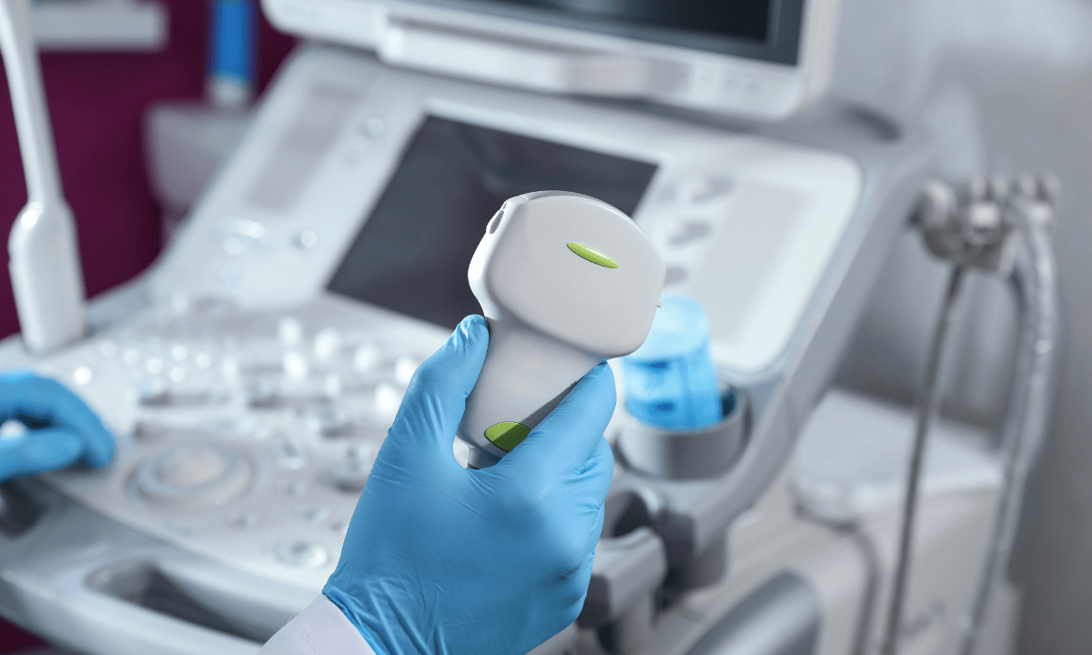
During the test, a healthcare provider will place a small device called a transducer against your chest. This device sends sound waves into your body that bounce back to create ultrasound images on a screen. These images help identify lung signs, such as air bronchograms, which are indicative of lung consolidation.
Lung ultrasound is used to identify several respiratory conditions, including the diagnosis of pneumothorax, pneumonia, interstitial syndrome, acute respiratory distress syndrome (ARDS), COPD exacerbation, and accumulation of fluid (pleural effusion). It often surpasses chest X-rays in accuracy, especially for detecting small pleural effusions.
One of the biggest advantages is that lung ultrasounds can be done right at your bedside, even if you're in the emergency room or intensive care unit. This feature means you can get fast answers without being moved to another department.
Your doctor might recommend a lung ultrasound if you have chest pain, difficulty breathing, or after certain procedures to make sure everything looks normal.
Why do doctors order a lung ultrasound?
Doctors may order a lung ultrasonography for various reasons:
For diagnosis of respiratory conditions:
Detecting fluid in the lungs (pleural effusion)
Diagnosing pneumonia or lung infections
Assessing lung collapse (pneumothorax)
Evaluating pulmonary oedema
Assessing COPD exacerbations or asthma complications
Monitoring certain lung diseases or conditions It’s particularly useful when a quick, non-invasive image of the lungs is needed, especially in emergency situations.
For procedural guidance:
Guiding thoracentesis (removing fluid from around the lungs)
Assisting with pleural biopsies
Biopsy: It may also provide guidance for performing biopsies of your lungs or the chest wall.
For monitoring and assessment:
Tracking treatment response for your lung conditions.
Evaluating unexplained shortness of breath.
Assessing chest trauma to check for complications.
Monitoring critically ill patients in the ICU.
To take charge of your health, book an appointment with Thomson Medical to meet our medical specialists and discuss a personalised treatment plan.
How do you prepare for a lung ultrasound?
Lung ultrasounds generally require little preparation. No special preparationis typically needed. However, before starting an ultrasound, please note that you:
Can eat and drink normally.
Should continue taking your regular medications.
Should wear loose, comfortable clothing to allow straightforward access to your chest and sides.
Should remove jewellery from your chest that might interfere with the ultrasound.
Inform your healthcare provider if you are pregnant or suspect you might be pregnant.
What happens during a lung ultrasound?

During a lung ultrasound:
You might lie on your back or side or sit up during the ultrasound. This depends on what the doctor needs to see.
A small amount of gel will be put on your chest. It helps the ultrasound probe send sound waves.
The technician will press a small handheld device called a transducer against different areas of your chest. As it moves across your skin, the transducer sends sound waves into your body and captures the echoes that bounce back, creating real-time images on a nearby monitor.
The procedure typically lasts 20 to 30 minutes.
Results
After the ultrasound, the images are reviewed by a radiologist or doctor to interpret the findings. They can identify conditions such as
fluid buildup (pleural effusion)
lung collapse (pneumothorax)
infections (pneumonia)
other lung abnormalities.
The results are usually available shortly after the procedure, providing timely information for diagnosis and treatment planning.
Are there any risks to doing a lung ultrasound?
Lung ultrasound is considered a very safe procedure with no significant risks associated with it. Unlike X-rays or CT scans, it does not involve radiation, making it suitable for all age groups, including children and pregnant women.
The non-invasive nature of the procedure ensures minimal risk of complications.
However, you might experience some minor discomfort during the process:
There may be slight pressure from the ultrasound probe as it is moved across your chest.
The ultrasound gel applied to your skin might feel cold initially.
Overall, these minor discomforts are temporary and do not pose any long-term risks.
FAQ
What does lung ultrasound show?
A lung ultrasound can show:
Pleural effusion (fluid in the pleural cavity)
Pneumothorax (air in the pleural space)
Infections or inflammation in the lungs
Pulmonary oedema (fluid in the lung tissues)
Certain types of lung tumours (in combination with other tests)
What are the 10 signs of a lung ultrasound?
Some of the signs that can be identified on a lung ultrasound include:
B-lines: indicative of pulmonary oedema (that means it suggest that the interstitial fluid in the lungs are present)
A-lines: normal lung pattern (suggests healthy lungs)
Consolidation: suggests pneumonia or infection
Effusion: fluid in the pleural space (pleural effusion)
Lung sliding: normal movement between the lung and pleura
Absence of lung sliding: It is a main indicator of pneumothorax
Comet tail artifact: suggests small pleural effusion
Shred sign: associated with pneumonia
Pleural thickening: can indicate chronic inflammation or malignancy
Subpleural consolidation: seen in conditions like pneumonia or infarcts
Can ultrasound detect water in the lungs?
Yes, ultrasound can detect water or fluid in the lungs, especially in the pleural space (pleural effusion). It can also help identify pulmonary oedema, which is fluid accumulation within the lung tissue.
How accurate is a lung ultrasound?
Lung ultrasound is generally considered highly accurate, especially for detecting pleural effusion, pneumothorax, and pulmonary edema. Its accuracy depends on the experience of the operator, but it is a valuable diagnostic tool, often used in emergency or critical care settings.
Can an ultrasound detect lung tumours?
Ultrasound may detect masses or abnormal growths in the lung, but it’s not the most definitive tool for diagnosing lung tumors. Advanced imaging techniques, like CT scans or MRIs, are typically used to assess lung tumours in greater detail. However, ultrasound can sometimes provide useful initial information when combined with other diagnostic methods.
What are the benefits of a lung ultrasound?
Lung ultrasound offers numerous benefits, particularly in critical care settings. As a point-of-care ultrasound (POCUS), it enables healthcare providers to perform bedside scanning, allowing for immediate assessment without the need to transport patients. This non-invasive ultrasound imaging technique is highly effective in detecting conditions such as pleural effusion and pneumonia, accurately identifying amounts of fluid in the pleural space. Additionally, unlike CT imaging, lung ultrasound does not expose patients to radiation, making it a safer option for ongoing evaluations in emergency and intensive care environments.
The information provided is intended for general guidance only and should not be considered medical advice. For personalised recommendations based on your medical conditions, request an appointment with Thomson Medical.
For more information, contact us:
Thomson Specialists Paragon (Health Screening)
- Mon - Fri: 8.30am - 5.30pm
- Sat: 8.30am - 12.30pm
Call: 6735 0300
Request a Health Screening