
What is a vascular ultrasound scan?
Vascular ultrasound is a non-invasive diagnostic test performed by trained sonographers. It uses high-frequency sound waves to show blood flow and the structure of blood vessels. Doctors use this diagnostic test to check arteries and veins in the neck, arms, legs, and abdomen.
This imaging test is often performed alongside a Doppler ultrasound study to monitor blood flow to organs and measure its movement, giving important details about your blood circulation. A vascular ultrasound can also help the medical professionals to detect various vascular conditions, including:
Blockages to blood flow and narrowing of vessels
Blood clots
Aneurysms (vessel bulging)
Other vascular abnormalities
Ultrasound is safe because it doesn't use ionising radiation. It has no known harmful effects and shows soft tissues that x-rays can't capture. This makes it a very safe diagnostic tool, without the risks of radiation. Patients usually have no side effects and can return to their normal activities right after the procedure.
When would I need a vascular ultrasound exam?
Your healthcare provider may recommend a vascular ultrasound if you have symptoms of blood vessel issues, such as:
Pain or swelling in the legs
Unexplained bruising or varicose veins
Symptoms of a stroke or a ministroke (transient ischaemic attack)
Poor circulation (cold or numb limbs)
The procedure helps your doctor detect various vascular conditions, including:
Atherosclerosis (hardening of the arteries)
Blood clots
Carotid artery disease
Chronic venous insufficiency (a condition where the veins in the legs are unable to properly pump blood back to the heart)
Deep vein thrombosis (a blood clot that forms in the deep veins in the body, usually in the legs)
Extracranial carotid artery aneurysm (a bulge in one of the arteries supplying blood to the brain)
Peripheral artery disease (PAD)
Varicose veins
Other vascular diseases
Additionally, a vascular ultrasound test is also used by your healthcare provider to assess an individual’s suitability for procedures such as angioplasty. It is also often used to monitor healing after surgeries or treatments to ensure the blood vessels are functioning properly.
If you’re experiencing any of the symptoms mentioned above, request an appointment with Thomson Medical. Our specialists can help to diagnose your condition, including performing a vascular ultrasound scan to determine the underlying causes and provide personalised care.
How do I prepare for a vascular ultrasound?
Preparation is usually minimal, and specific instructions depend on the type of vascular ultrasound you are having. Here are the common guidelines to follow:
Fasting
You may need to fast for a few hours before the exam, particularly if the ultrasound will examine your carotid arteries or abdominal blood vessels.
Clothing
Wear loose, comfortable clothing that allows easy access to the area being examined. You might need to change into a hospital gown for the test.
Medications
In most cases, you can continue taking your prescribed medications. However, it’s essential to inform your healthcare provider about any medications you are currently taking.
Always adhere to any specific instructions provided by your healthcare professional before the exam to ensure accurate results.
How does vascular ultrasound work?
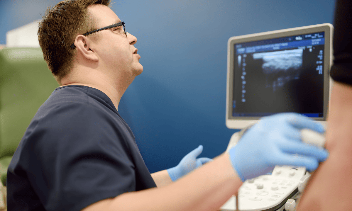
Vascular ultrasound uses a handheld device called a transducer that emits high-frequency sound waves. These sound waves travel through your soft tissues and blood vessels when applied to your skin over the area under examination.
As they encounter different types of tissue, the sound waves create echoes that bounce back to the transducer. The ultrasound machine then processes these echoes and creates images or videos of your blood vessels. This technology allows your doctor to visualise blood flow in real-time and evaluate the structure and function of your arteries, veins, and other vascular structures.
What will I experience during and after the procedure?
During the procedure
You'll be positioned on an examination table, and a clear, water-based gel will be applied to the area being studied. This gel helps the transducer maintain good contact with your skin and eliminates air pockets that could interfere with the sound waves. As the sonographer moves the transducer across your skin, you may feel slight pressure but typically no pain.
The examination usually takes between 30-60 minutes to complete, depending on how many blood vessels need to be evaluated and the complexity of your case.
After the procedure
Once the ultrasound is finished, the gel will be wiped off your skin and you can immediately return to your normal activities.
What do the test results reveal?
Vascular ultrasound provides healthcare providers with detailed imagery that illustrates patterns of blood flow through your body. The test supplies vital information in relation to:
The blood flow rate through your blood vessels
Anything that can block it, such as plaques or blood clots
Regions of narrowing of blood vessels
The overall structure and function of veins and arteries
This complete image enables your doctor to accurately diagnose conditions that impact your blood vessels and determine the most effective treatment for your situation.
Always follow preparation guidelines and discuss any concerns with your doctor to ensure the most accurate results. Request an appointment with Thomson Medical to find out if a vascular ultrasound is the right imaging test for you.
Does vascular ultrasound have any risks?
Vascular ultrasound examination is considered a safe medical procedure with no known risks. It uses sound waves instead of radiation, so there's no exposure to harmful rays like with X-rays or CT scans.
Most patients experience no discomfort during or after the examination. However, in rare cases, you might develop mild, temporary skin irritation from the gel applied during the procedure, particularly those with sensitive skin.
The gentle pressure applied by the transducer during the examination may cause a little discomfort if you have a sensitive area that is being examined, but this sensation is brief and disappears quickly after the procedure.
FAQ
What can I expect from a vascular ultrasound of the leg?
A vascular ultrasound of the leg is often performed to assess blood flow and detect conditions like deep vein thrombosis (DVT), peripheral artery disease (PAD), or other vascular issues in the leg. Here’s what to expect:
Positioning
You will be asked to lie down on an exam table, and your leg may be raised or positioned in different ways for better access to the arteries and veins.
Gel application
A gel will be applied to your leg to help the ultrasound probe make good contact with your skin. The gel allows sound waves to pass through more effectively.
Transducer use
The technician or doctor will move a small device (transducer) over your skin, which emits sound waves and captures the returning echo waves to create an ultrasound image of the flow of blood in your veins and arteries.
Duration
The procedure usually lasts about 30 to 60 minutes.
What you may feel
The procedure is generally painless. You may feel slight pressure when the technician presses the transducer against your leg, but it shouldn’t be uncomfortable.
What is the difference between a cardiac ultrasound and a vascular ultrasound?
A cardiac ultrasound (also known as an echocardiogram) and a vascular ultrasound both use sound waves to generate images, but they focus on different parts of the body:
Cardiac ultrasound
The test focuses on the heart and its functions.
It helps to assess the heart’s chambers, valves, and blood flow and can detect heart conditions like heart failure, valve issues, and congenital heart defects.
Vascular ultrasound
The test focuses on the blood vessels throughout the body (arteries and veins).
It helps to detect issues like blockages, blood clots, or narrowing of blood vessels in areas like the legs, neck (carotid arteries), or abdomen.
Can ultrasound detect blocked arteries?
Yes, ultrasound scanning can identify blocked arteries. Doppler ultrasound is particularly useful for this. It evaluates blood flow and detects any narrowing or blockages in the arteries.
What is the best treatment for blocked arteries in the legs?
The treatment for blocked arteries in the legs depends on the severity of the condition and is usually caused by peripheral artery disease (PAD):
Lifestyle changes
Exercise
Regular walking can make a big difference! Even short walks can help improve blood flow to your legs and reduce pain or discomfort.
Diet
Adding more fruits, vegetables, and whole grains to your meals while cutting back on fatty and processed foods can help stop blockages from getting worse.
Smoking cessation
If you smoke, stopping is one of the most important steps you can take. Smoking damages blood vessels and makes PAD symptoms much worse.
Medications that might help
Antiplatelet drugs (like aspirin or clopidogrel) to prevent blood clots.
Cholesterol-lowering medications to reduce plaque buildup.
Blood pressure medications that can help you manage high blood pressure.
Non-surgical procedures
Angioplasty
A procedure where a balloon is inserted into the blocked artery to widen it.
Stenting
A small mesh tube is placed in the artery to keep it open.
Atherectomy
Removal of the plaque from the artery using a catheter.
Surgical treatment:
Bypass surgery
A surgeon creates a new path for blood to flow around the blocked artery, often using a vein or synthetic graft.
The best treatment plan depends on the severity of the blockage, symptoms, and overall health, and it is usually tailored by a vascular specialist.
What are the different types of a vascular ultrasound?
Some common types of vascular ultrasound are:
Carotid ultrasound (Carotid duplex)
This test evaluates blood flow through the carotid arteries in the neck to detect blockages or narrowing caused by plaque buildup, which can increase the risk of stroke.
Peripheral venous ultrasound (DVT)
This test focuses on detecting blood clots in veins, particularly in the legs, and is commonly used to diagnose deep vein thrombosis (DVT).
Peripheral arterial ultrasound (ABI)
This test assesses blood flow in the arteries of the arms and legs, often used to diagnose peripheral artery disease (PAD).
Duplex ultrasound
This test evaluates blood flow in both arteries and veins throughout the body. It provides information on blood speed, identifies blockages, and assesses the overall condition of the vascular system.
Renal artery ultrasound
This imaging evaluates the arteries supplying blood to the kidneys, helping to detect narrowing or blockages that could lead to renal dysfunction or hypertension.
When will I receive my ultrasound results?
When will I receive my ultrasound results? The results of a vascular ultrasound are typically analysed by a radiologist or vascular specialist. After the evaluation is complete, the findings are sent to the healthcare provider who ordered the test. Typically, patients can expect to receive their results within a few days after the procedure. At this time, your healthcare provider will discuss the implications and any necessary follow-up actions based on the findings.
The information provided is intended for general guidance only and should not be considered medical advice. For personalised recommendations based on your medical conditions, request an appointment with Thomson Medical.
For more information, contact us:
Thomson Specialists Paragon (Health Screening)
- Mon - Fri: 8.30am - 5.30pm
- Sat: 8.30am - 12.30pm
Call: 6735 0300
Request a Health Screening